Monday, October 2, 2023

Disordered proteins: the next frontier in therapeutics?


Howard Stone at Princeton, Ozgur Sahin at Columbia, and their colleagues have written a wonderfully imaginative review of what they call “hydration solids” (Nature 619, 500; 2023 - here). By this I don’t mean to imply anything fantastical about it – I simply like this way of framing the notion that hydration water can play an important structural and mechanical role in some hygroscopic biological materials. The idea here is that the mechanical properties are governed by the hydration forces operative within a fluid-filled porous elastic medium. The notion is motivated and explored by studying the mechanical behaviour of bacterial spores, but the same principles might apply to wood, pollen grains, keratinous materials and silk. “Such ‘hydration solids’, which can exchange their essential constituent water with the environment and have it flow through the material, are potentially abundant in the environment”, they write.


There is a fascinating paper in PRL from Chunyi Zhang (Mike Klein’s group) at Temple University in Philadelphia that investigates why the dielectric permittivity of salt water can actually decrease as more salt is added (C. Zhang et al., Phys. Rev. Lett. 131, 076801; 2023 - here). Using a deep neural network trained on the results of density functional theory, the authors show that this is not some kind of saturation effect but arises because of the way the ionic hydration shells disrupt the hydrogen-bonded network of the water and thereby suppress the collective response to electric fields.


It has been recognized at least since the early 1970s (and explored by the late, great Jack Dunitz) that changes in enthalpy and in entropy of associations between biomolecules (such as receptor-ligand pairings), due for example to small changes in molecular structure, seem often to compensate for one another so as to entail little change in the Gibbs free energy of binding. Why this is so has been much debated. An intuitive explanation is that a more favourable enthalpic contribution to binding creates a corresponding decrease in conformational freedom and thus a loss in entropy. But consideration of that balance must also take into account changes in hydration due to reorganization of the local hydrogen-bonded network. The Whitesides group (Breiten et al., JACS  135, 15579; 2013) has argued that water reorganization is in fact the key source of enthalpy-entropy compensation. That idea is examined, and ultimately supported, in a study by Shensheng Chen and Zhen-Gang Wang at Caltech (J. Phys. Chem. B 127, 6825; 2023 - here), using MD simulations of model charged polymers. They find that the hydrophobic interactions resulting from reorganization of hydration water show temperature dependencies that can account for the close correlation between the deltaH and TdeltaS terms. The effects of electrostatic interactions and polymer conformational changes are, in comparison, minor. Water is, it seems, in control.

Liquid-liquid phase separation (LLPS) has become a vibrant topic in cell biology now that it’s clear cells make use of it in a variety of ways and circumstances for partitioning and sequestering biomolecules for purposes ranging from gene regulation to RNA splicing to stress responses. The globular droplets – condensates – formed in this process have a higher density than the surrounding cell fluid, but it’s still not really understood what characteristics of biomolecules promote this new phase. That understanding could be useful for being able to control the phase separation process for possible therapeutic purposes – or indeed for designing peptides to prevent pathogenic aggregation. The condensates are not biomolecular complexes in any real sense – the binding forces between the components seem to be rather weak and indiscriminate, and condensates typically contain proteins with some degree of disorder (intrinsically disordered proteins, IDPs), which tend to be promiscuous in their interactions. Debasis Saha and Biman Jana of the Indian Association for the Cultivation of Science in Kolkata have used MD simulations to investigate the factors that govern the interactions of model peptides, leading to dimerization, to try to get some handle on what is going on (J. Phys. Chem. B 127, 6656; 2023 - here). They consider the effect of charged residues such as arginine, as well as differing amounts of hydrophobicity in the chains, in altering the free energy surface for dimerization. For both positively and negatively charged peptides, the solvation water seems to play an important role, and is the dominant influence in the latter case. The upshot is that, if dimerization adequately reflects the condensate formation process, negatively charged peptides seem more likely to stay in the dilute phase.

Benjamin Schuler at Chicago and colleagues look at a similar issue: the interaction between the highly positively charged histone linker H1 and the highly negatively charged prothymosin α (ProTα) which acts as a “chaperone” that can help H1 disassociate from the histone (A. Chowdhury et al., PNAS 120, e2304036120; 2023 - here). Both are IDPs. They find that in this case there is a large entropic contribution to the binding coming from the release of counterions – a consideration that they expect to be most generally applicable to bio-polyelectrolyte interactions.  

Meanwhile, in a paper in bioRxiv [here], Saumyak Mukherjeee and Lars Schäfer at Bochum have looked at the thermodynamic driving forces that govern the formation of condensates from proteins with intrinsically disordered domains. Aggregation into the dense phase will involve changes in enthalpy and entropy due both to direct protein interactions and to changes in solvation. The authors conclude from MD simulations that in this case the most important factors are protein interaction enthalpy and the entropic effects of water release from the protein hydration shell into the bulk.

It seems likely that unravelling these factors governing IDP associations of various sorts will ultimately be valuable as we seek to make pharmaceutical interventions in molecular interactions of this kind that seem connected to disease (such as Alzheimer’s). Christine Lim at Cambridge and colleagues have developed a bioinformatics platform for identifying proteins involved in LLPS that seem to be potential therapeutic targets (C. M. Lim et al., PNAS 120, e2300215120; 2023 - here). They test it by looking at the in vitro phase behaviour of three targets that their scheme identifies. I don’t think it is too much to suggest that this points towards something of a new paradigm for therapeutics, in which the goal is not to develop some inhibitor that might compete with a protein’s normal ligand but rather to engineer collective and less selective interactions at a larger scale.

I’m intrigued by a paper in press in J. General Physiology (preprint here) by Alan Kay at the University of Iowa and Gerald Manning at Rutgers, arguing that what drives osmosis is still not fully understood and that the mechanism proposed by Peter Debye in 1923 is in fact the right one, despite being now largely forgotten. It is one thing to explain osmotic flow thermodynamically in terms of differences in chemical potential (the textbook account), but another to explain what actually drives the directional transport of water molecules. Manning and Kay say that diffusion alone is not able to account for the osmotic flux, which arises instead because of differential repulsive forces between the solute molecules and the two interfaces of the semipermeable membrane. This produces the equivalent of a hydrostatic pressure difference that drives the flux. I’m certainly not qualified to assess whether this revision of the textbook explanation is valid, but the history of the Debye model given in the paper is surely interesting in its own right.  

There is, admittedly, not much biology going on in the deep mantle of the Earth with temperatures of 1,000-2,000 K and pressures of up to 22 GPa. All the same, such conditions pose quite a test of molecular-dynamics water potential functions, which Roberto Car at Princeton, Giulia Galli at Chicago, and colleagues have put through their paces (C. Zhang et al., J. Phys. Chem B 127, 7011; 2023 - here). They set out to calculate the thermal conductivity of water in these conditions, using a potential found by fitting to density-functional theory using a deep-learning algorithm. The conductivity varies only slightly with temperature, decreasing from its ambient value, but is much more strongly (and positively) correlated with density. The heat transport of water at high P and T could have important implications for processes in the interiors of gas-giant planets.

Monday, July 24, 2023

It's back!


Back by popular demand” always seemed a self-congratulatory phrase, but in this case it really is the case that I’m reviving this blog because so many people have said they found it helpful. I’m touched by that, and also grateful to be given the motivation to continue pursuing this endlessly fascinating topic. So here we go.


One of the things that led me to let the blog languish was that there were so many other areas in which I have been striving to deepen my knowledge over the past several years. Happily, at least one of these remains relevant to the topic at hand: I have been delving deeper into the thickets of molecular and cell biology for my latest book, How Life Works, which is published in the autumn/fall of 2023. Among many other things, this looks at issues such as the roles of disordered proteins and of biomolecular condensates, in which issues of solvation are clearly important.


As if to underscore that, I am kicking off this post with a fascinating paper in Nature by Benjamin Schuler at Zurich and colleagues [N. Galvanetto et al., Nature 10.1038/s41586-023-06329-5; 2023], which looks at biomolecular dynamics within such a dense condensate. I probably don’t need now to say much about the importance of condensates in cell biology; as Schuler and colleagues say, these dense but loose associations of proteins and nucleic acids “play a key role in cellular processes operating at different scales, such as ribosome assembly, RNA splicing, stress response, mitosis and chromatin organization, and they are involved in a range of diseases” (and also, I’d add, in gene regulation). Their significance was confirmed by the fact that Tony Hyman and Cliff Brangwynne, who did much of the early work to bring them to wider attention, were awarded last year’s Breakthrough Prize in the life sciences. As Schuler et al. say, intrinsically disordered proteins seem often to play a key part in the formation of condensates, thanks to their rather promiscuous binding capacity.


The dense fluid of these phase-separated blobs can have a greatly enhanced local concentration of constituents relative to the bulk cytoplasm, and indeed this is a central feature of their function: they help to concentrate particular biomolecular species to enable repeated, low-affinity encounters of the kind that seem important for e.g. gene regulation or splicing. That density, however, results in a water viscosity that can be several orders of magnitude greater than it is in the bulk. Schuler and colleagues use single-molecule spectroscopy and MD simulations to examine the consequences of this for the dynamics of proteins. They study in vitro proxies for in vivo condensates, consisting of droplets (coacervates – I’m interested to see the return of that word from the days of the “prebiotic” speculations of Sidney Fox!) made from two highly and oppositely charged IDPs, both of which are known to influence chromatin condensation and transcriptional regulation.


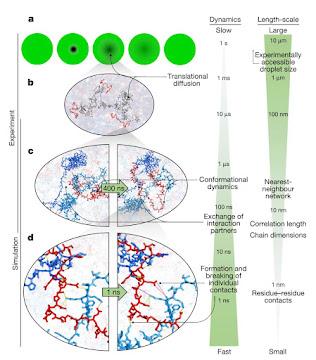
The result is that, despite a 300-fold increase in water viscosity in the droplets and the formation of a dense network of indiscriminately associated proteins, leading to drastically slowed translational motion, the local conformational dynamics of the chains that influence residue-residue contacts remain fast, happening on pico- to nanosecond timescales. The conclusions are nicely summarized in this image:


As the authors say: “The behaviour we observe is an example of the subtle balance of

intermolecular interactions in biomolecular phase separation. On the one hand, the interactions must be strong enough for the formation of stable condensates; on the other hand, they need to be sufficiently weak to enable translational diffusion and liquid-like dynamics within the dense phase and molecular exchange across the phase boundary— processes that are essential for function, such as biochemical reactions occurring in condensates.” I have to say that this doesn’t entirely surprise me, as it seems to reflect in a more extreme way what seems to happen in cells more generally: the cytoplasm can look somewhat gel-like at mesoscales, and translational diffusion can be anomalous, while at the molecular scale dynamics are not so different from those in dilute solution. Of course, the usual caveats about in vitro studies – which have been stressed in particular for studies of condensates (see e.g here) – apply too. But this is a really nice study, which to my mind illustrates the possibility of a separation of dynamical timescales that makes life possible: the benefits of forming condensates do not need to come at the expense of compromising the local dynamics needed for biomolecular function.


Needless to say, I have from time to time seen some wonderful papers touching on water in biology come and go in the time between my last blog and now, and I fear these are lost now to being documented here. But there are a few recent ones that I still have to hand. There has been a particularly vigorous debate in the past year or two on the issue of the putative liquid-liquid phase transition in pure water. The evidence for this continues to accumulate, and looks now to be rather strong – although that clinching proof remains elusive. This paper [Science 359, 1127; 2018] by Sander Woutersen in Amsterdam – including the late and much missed Austen Angell – reported a direct sighting of such a transition in an aqueous solution of hydrazinium trifluoroacetate, which could be deeply supercooled to 140 K. And work by Yoshiharu Suzuki in Tsukuba adds to that suggestive evidence with a study [PNAS 119, e2113411119; 2022] of supercooled aqueous trehalose, which reports a direct signature of a first-order liquid-liquid transition. You can see some responses to the work here. Meanwhile, Nguyen Vinh and colleagues at Virginia Tech have performed terahertz measurements of water over a wide temperature range, down to the deeply supercooled state, which they say can be interpreted it terms of two liquid forms with different transition temperatures.  


I am (not so?) secretly delighted that my PhD supervisor Bob Evans at Bristol decided some time ago to delve into water phase behaviour, having warned me (wisely!) to steer clear of any liquid so anomalous. Here [J. Chem. Phys. 158, 034508; 2023] Bob, Mary Coe and Nigel Wilding suggest that the well known density depletion and enhanced fluctuations evident in water near hydrophobic surfaces be interpreted as the remnant of a critical surface (drying, or as some call it, dewetting) phase transition. In other words – and this seems to me quite significant – there is in a sense nothing unusual about water in this respect, and no need to invoke any specialness about its liquid-state structure. Rather, this is well-understood statistical physics at work. I notice that this was the topic of Mary’s 2021 PhD thesis: “Hydrophobicity across length scales: The role of surface criticality” – which I’d love to see.


I can’t touch on that topic without remembering that for my last post in January 2018, I seem not yet to have caught up with the news of the death in April 2017 of David Chandler, a giant in the field of liquid-state theory, as well as statistical mechanics more broadly. David’s work with John Weeks and Ka Lum on the hydrophobic interaction [JPCB 103, 4570; 1999] has of course been one of the most influential contributions to this topic since it was published in 1999. David’s work has certainly been a huge influence on my thinking in this field – and indeed that influence went right back to my work with Bob in the mid-1980s. David was always generous but rigorous, and phenomenally insightful, and his absence is deeply felt.


Francesco Paesani of UCSD has let me know about the new water potential he and his colleagues have developed from first-principles quantum simulations, which is able to capture the phase diagram very realistically [Bore & Paesani, Nat. Commun. 14, 3349; 2023] – see also the improvements in Zhu et al., J. Chem. Theory Comput. 19, 3551-3566; 2023]. Francesco has already been applying it to a variety of real-world problems, including the hydration of a simple model of the protein backbone (N-methylacetamide) [Zhou et al., J. Chem. Theory Comput. 19, 4308; 2023].


I just noticed this interesting article [A. Katsnelson, ACS Cent. Sci. 10.1021/acscentsci.3c00803; 2023] on “artificial saliva” and synthetic mucins. I knew nothing about such work, which sound like rich ground for exploring hydration issues.


I can’t end this first post without mentioning the ongoing story of “life (or not) in the clouds of Venus”. This intriguing idea was first put forward by Sara Seager and her colleagues following the apparent identification [J. S. Greaves et al., Nat. Astron. 5, 655; 2021] of phosphine – for which no abiotic natural source is known – in the Venusian atmosphere. Much of the immediate debate (aside from that about how secure the signature of phosphine really was, a discussion that is still ongoing) centred on the extreme acidity of the cloud droplets. But I was more concerned about how low the water activity in the droplets must be – we know that no life on Earth has been seen for water activities below 0.585, a fact that I am sure is related to the minimal hydration requirements for functional biomolecules. When I mentioned this to microbiologist John Hallsworth at Queens in Belfast, he convened a team of experts who made calculations to estimate the water activity in the Venusian clouds and a variety of other planetary environments, revealing that on Venus this is indeed orders of magnitude too low to permit terrestrial-type life given what we currently know about it [J. E. Hallsworth et al., Nat. Astron. 5, 665; 2021]. Sara and her coworkers have now suggested [W. Bains et al., Astrobiol. 10.1089/ast.2022.0113; 2023] possible ways around this problem, which in the end amount to having to posit life unlike that on Earth – a possibility we recognized in our paper. To my mind, this could usefully sharpen the discussion about what “habitable” can and should mean in astrobiology, as I explore here. I welcome anything that prompts deeper, quantitative thinking about the conditions needed to support something we might reasonably call life in extraterrestrial environments. But I would argue that such discussions need to go beyond “well, X can also act as a hydrogen-bonding solvent”, and should take into consideration just how subtle water’s roles are in terrestrial biochemistry, and how much the “specialness” of water might matter in this regard. Given the richness of emerging knowledge about extraterrestrial environments, and the prospects of much more to come through planned missions (e.g. to Enceladus and Titan) and JWST observations, I’d love to see this issue engaged with head-on by e.g. ESA or NASA.   


Well, there we are: I’m open for business again. So do feel free to send things my way for inclusion; I’d be delighted to hear about them.  

Wednesday, January 24, 2018

Hydration as a design element in biomaterials

Given what is known about water’s active role in the structure, dynamics and function of biomolecules, can it be used as a design element in synthetic molecular and nanoscale structures derived from them, which might have more amenable levels of complexity? Sam Stupp at Northwestern and colleagues ask this question in relation to the amphiphilic peptide-based nano-architectures they have been developing. They have used Overhauser dynamic nuclear polarization relaxometry to characterize the hydration of such a peptide nanofibre [J. H. Ortony et al., JACS 139, 8915; 2017 – paper here]. They find a wide range of water dyamics, from rapid translational diffusion within the hydrophobic interior to near-immobilization of water on the hydrophilic surface. Water further from the core and close to the inner peptide domain is slowed relative to the bulk, and more so for fibres in the gelled than in the solution state. The same applies to water close to the (charged) nanofibre surface, which may be slowed to a degree comparable to that of water confined within protein cavities. MD simulations echo these findings. Sam and colleagues suggest that the surface water might play an active role in gelation and interactions with biomolecules, and in consequence that “water dynamics not only play an important role in the function of nanostructured biomaterials, but… may also be tunable in soft matter at molecular length scales to optimize performance for a variety of biomedical applications.”

In a similar spirit, Geraldine Richmond at Oregon and coworkers use vibrational sum frequency spectroscopy to look at water orientational ordering close to the surface of nanoemulsion aggregates: micelles and reverse micelles [J. K. Hensel et al., PNAS 114, 13351; 2017 – paper here]. Water seems to be electrostatically ordered by the surfactant head groups and their counterions at the interface.


Proposed orientation of water at surfactant (AOT)-counterion surfaces, both for the curved surfaces of micelles and the planar surfaces of lamellae.

How exactly does one quantify water order or disorder? Various measures have been suggested; Fabio Sterpone of the Université Paris Diderot and colleagues suggest that it might be sought in the connectivity of the hydrogen-bond network (O. Rahaman et al., JPCB 121, 6792; 2017 – paper here). They have looked at this measure in simulations of the protein (here lysozyme) dynamical transition at around 240 K. There is a fairly abrupt change in the connectivity at the transition, in that the degree of percolation is constant below about 220-240 K but decreases with temperature above it. A closer analysis shows that at 240 K the hydration water network begins to sample a larger number of configuration states, corresponding to an abrupt increase in configurational entropy. Fabio and colleagues interpret these findings in terms of the hydration water acting as an ‘entropic reservoir’ that, above the dynamical transition, enables the protein to undergo configurational changes.


Probability distribution of hydration-water percolation propensity for lysozyme as a function of temperature.

Not strictly water in biology, but Daniel Munoz-Santiburcio and Dominik Marx in Bochum describe ab initio simulations of proton transport in alkaline water films confined between the layers of the mineral mackinawite [Nat. Commun. 7, 12625; 2016 – paper here]. They find that orientation and coordination of hydroxide can alter the hydrogen-bond network in ways that make transport of protons and mobility of negative-charge defects sensitive to the width of the confining space: different structures in the layered water films support qualitatively different transport mechanisms. Daniel and Dominik conclude that “it should be possible to rationally design different nanostructures that will allow for different charge transport rates of confined alkaline aqueous solutions.” Meanwhile, in another paper they show that water within mackinawite layers has a boosted tendency to self-dissociate (Phys. Rev. Lett. 119, 056002; 2017 – paper here).

And in a third paper they consider the effect of water confinement (between mineral layers) on a whole set of chemical reactions, particularly those involved in peptide formation – which may serve as a model of prebiotic chemistry at nanoconfined mineral surfaces (Chem. Sci. 8, 3444; 2017 – paper here). Under these conditions both the energetics and the mechanisms of the reactions can be significantly altered relative to the bulk.

X-ray free electron lasers are already transforming structural biology with their ability to provide diffraction data on very small samples in some cases down to the single-molecule scale, at high time resolution for studying dynamical processes. Jessica Thomaston and coworkers have used the Japanese Spring-8 XFEL source to obtain a new structure of the influenza M2 proton channel under ambient conditions (J. L. Thomaston et al., PNAS 114, 13357; 2017 – paper here). They find that an ordered network of hydrogen-bonded water molecules spans the pore at pH5.5, providing a proton-conduction pathway and stabilizing the protonated His37 residue present in the intermediate open state of the channel. These ordered waters decrease in number as pH increases and the open state becomes less stable.


The room-temperature XFEL structures of the M2 channel at different pH. Red spheres show waters with full occupancy, light and dark blue with half-occupancy.

The idea has been around for some time that water might play an important role in protein (mis)folding into amyloid fibrils associated with neurodegenerative diseases. Mei Hong at MIT and colleagues have now used solid-state NMR to characterize the reservoirs of water within and around fibrils of the Alzheimer’s β-amyloid peptide Aβ-40 (T. Wang et al., JACS 139, 6242; 2017 – paper here). They find five distinct pools, ranging from surrounding bulk-like matrix water to mobile interfibrillar water channels and relatively immobile peptide-bound reservoirs. Exchange of water happens between the two dynamic pools on very long (second) timescales. The balance between the various pools is shifted in some mutant fibrils.


The five water pools in wild-type Aβ-40 amyloid fibrils.

Much attention has been given to the roles of hydration water in the binding of ligands by proteins, but less to the idea that solvation is also important for unbinding. Paolo Carloni at Jülich and colleagues have simulated the unbinding kinetics of an anti-inflammatory agent (a urea derivative) that binds to p38 MAP kinase (R. Casanovas et al., JACS 139, 4780; 2017 – paper here). They find that a rotation of the urea group during unbinding creates a more –solvent-exposed state in which the hydrophobic interactions between the t-butyl substituent of the ligand and a hydrophobic part of the cavity are weakened. Water molecules that enter into the cavity play an essential role in this step, forming hydrogen bonds to the urea group and mediating those with residues in the cavity.

An important role for water-mediated contacts is identified in the DNA binding of transcription factors of the ETS family by Gregory Poon and colleagues at Georgia State University (S. Xhani et al., JPCB 121, 2748; 2017 – paper here). One in particular, denoted PU.1, shows a strong osmotic sensitivity in its binding, which the researchers attribute to the water-mediated binding between a tyrosine residue and DNA. Mutation of this residue removes this sensitivity. In the homologue Ets-1, which lacks this sensitivity, it seems that the same Tyr residue, while apparently engaging in the same water-mediated contact in the bound state, actually plays a different role in binding, by influencing the local dynamics of the free protein. I guess this underscores once more how hard it is to identify general rules of thumb for how hydration structures affect biomolecular function and molecular recognition.


The same and not the same: these two water-mediated contacts between a Tyr residue and DNA in the homologous transcription factors PU.1 (top) and Ets-1 (bottom) actually play different roles in the molecular-recognition process.

Cytochtome c oxidase (CcO) pumps protons across membranes using the energy of dioxygen reduction. The mechanism seems to involve a change in hydration – a switch between ‘wet’ and ‘dry’ configurations – in an internal cavity that is connected to the proton’s exit channel. Qiang Cui and colleagues at the University of Wisconsin have studied that process using MD simulations (C. Y. Son et al., PNAS 114, E8830; 2017 – paper here). They show how the protonation state of one residue acts as a switch between the dry and wet states, and that in the wet state the pKa of a Glu residue is lowered to facilitate proton transfer. Here, then, is a functional role for the dry-wet switching of the cavity.

What about the proton channel itself in CcO? It’s not in fact yet clear what route it takes – three possible channels (labelled D, K and H) have been proposed. Vivek Sharma at the University of Helsinki and colleagues use simulations to show that the H channel alone doesn’t seem able to do the job, unless a buried histidine residue is protonated (which seems unlikely); otherwise there’s a gap that the transient water networks in the channel can’t span (V. Sharma et al., PNAS 114, E10339; 2017 – paper here).

Here’s a rather old paper that I only noticed recently. Molecular recognition in proteins is still often discussed in biochemistry textbooks in terms of the induced-fit model. But there’s more to it. An alternative (if not necessarily mutually exclusive) picture has been developing recently in which binding occurs preferentially for particular, perhaps weakly populated conformations in an ensemble of protein states, stabilizing that conformation. Oliver Lange at the MPI Göttingen and colleagues used NMR to characterize the conformational states of ubiquitin in solution, and their results offer support for this notion of conformational selection (O. F. Lange et al., Science 320, 1471; 2008 – paper here). This work strengthens the idea that protein dynamics – which of course are highly influenced by hydration and by solvent fluctuations – have a prominent role in their functionality, an issue I discussed briefly in a recent column in Chemistry World (here https://www.chemistryworld.com/opinion/snapshots-of-lifes-dancers/3008393.article). The Lange et al. work was nicely summarized in a Perspective article by David Boehr and Peter Wright at Scripps (Science 320, 1429; 2008 – paper here).

In an intriguing simulation study, Sridip Parui and Biman Jana at the Indian Association for the Cultivation of Science in Kolkata have looked at the hydrophobic interactions between two hydrocarbon molecules, and also two rod-like model hydrophobes, at low temperatures (240 K) (JPCB 121, 7016; 2017 – paper here). They find that there is a second solvent-separated minimum for both systems, roughly 1 nm apart. This corresponds to weaker hydrophobic interactions between the solutes, due to stronger water-water interactions. The authors suggest that such a state could play a role in cold denaturation of proteins.

The effect of osmolytes on the denaturation of a model protein (stem bromelain) is considered in detail by Pannuru Venkatesu of the University of Delhi and coworkers (A. Rani et al., JPBC 121, 6456; 2017 – paper here). They use a whole battery of experimental techniques (fluorescence, UV-Vis and circular dichroism spectroscopy and dynamic light scattering), along with MD simulations, to study the stabilizing effect of a series of osmolytes (proline, betaine, arginine, sarcosine) against thermal denaturation. In all cases the effects seem to be caused by direct interactions. Betaine, sarcosine and arginine all interact with the protein via hydrogen-bonding. Proline, meanwhile, which confers the greatest stabilizing effect, binds in the protein’s active site.

Michael Feig at Michigan State and others have published a nice review article on biomeolecular crowding (M. Geig et al., JPCB 121, 8009; 2017 – paper here). This considers the effects of crowding on molecular diffusion, conformation and dynamics, but also on the solvent, which – as the authors say – can have altered structure, dynamics and dielectric response in typical crowded geometries.


A typical crowded environment in the cell.

Coralie Pasquier at the University of Lund and coworkers have studied the effect of electrolytes with multivalent (trivalent yttrium) ions on protein-protein interactions, using MD and MC simulations (C. Pasquier et al., JPCB 121, 3000; 2017 – paper here). The Y ions bind to the surface of the proteins (human serum albumin), but the consequences are complex. In a coarse-grained model, increasing Y3+ concentration could increase protein repulsion at low ionic strength but increase it at high ionic strength. The first situation is due to double-layer effects, the second is a Coulombic repulsion due to high charging of the protein surface. These interactions are water-mediated, and screened out by addition of NaCl, resulting in protein attraction. At intermediate concentrations of YCl3 there is also a net attraction between the proteins, due to ion-ion correlations.

Takeshi Yamada of CROSS in Naka, Japan, and coworkers have used quasi-elastic neutron scattering to look at the dynamics of water sandwiched between phospholipid bilayers (T. Yamada et al., JPCB 121, 8322; 2017 – paper here). They see three distinct populations of water molecules: free and almost bulk-like, loosely bound and tightly bound to the phospholipid head groups.

Concentrated ionic solutions aren’t so commonly encountered in biology, but they are important in some technologies, such as rechargeable aqueous batteries. Water is known to have slowed rotation in such solutions, and Wei Zhuang of the Fujian Institute of Research on the Structure of Matter and coworkers suggest why (Q. Zhang et al., PNAS 114, 1123; 2017 – paper here). Their simulations suggest that the key contribution comes from a coupling of the slow, collective component of rotation with ion clusters, rather than from faster single-molecule motions. They say that there are similarities with water rotations near large biomolecules.

Do we finally have an accurate, predictive, tractable ab initio model of water? Mohan Chen at Temple University and collaborators think so (M. Chen et al., PNAS 114, 10846; 2017 – paper here). They call their model the strongly constrained and appropriately normed (SCAN) density functional, and say that it captures many of the structural, dynamic and electronic properties of liquid water as well as the density difference with ice Ih.

Talking of such things, Tomotaka Oroguchi and Masayoshi Nakasako of Keio University say that to accurately simulate the directionality of hydrogen bonds from donor atoms in hydrophilic amino acid residues, one needs to include in the force fields off-atom charge sites that mimic the lone-pair electrons (Sci. Rep. 7, 15859; 2017 – paper here).

Monday, June 19, 2017

Chiral water in DNA's hydration shell

In a clever study of DNA hydration using SFG spectroscopy, Poul Petersen and his coworkers have found that the chiral spine of hydration in the minor groove, inferred from oxygen locations for hydrated crystalline DNA by Dickerson and collaborators in the 1980s, exists also in aqueous solution under ambient conditions, and entails orientational ordering of the hydrogen bonds in the single-file water chain that fits into this narrow groove (M. L. McDermott et al., ACS Centr. Sci. 10.1021/acscentsci.7b00100; 2017 – paper here). I wrote a news story for Chemistry World on this work (here).

I applaud the ambition of Modesto Orozco of the Barcelona Institute of Science and Technology and colleagues in writing a paper called “The multiple roles of waters in protein solvation” (A. Hospital et al., JPCB 121, 3636; 2017 – paper here). There’s a title guaranteed to say to me “Read this now!” And the ambition continues in the extent of the systems they investigate with MD: a range of proteins, at a range of temperatures, some denatured, some with crowding agents, some with high concentrations of urea. They say that the results illustrate “the dramatic plasticity of water, and its chameleonic ability to stabilize proteins under a variety of conditions”, which seems a fair way to summarize the matter. I’m not sure I see any surprises here, and the denaturant effects of urea are discussed with something of a “water structure” flavour, but it’s a kind of snapshot of the sorts of things hydration water gets up to.

A more specific study of protein hydration dynamics is described by Dongping Zhong and colleagues at Ohio State University, who use tryptophan as the reporter group to characterize the dynamics at 17 sites on the surface of the β-barrel protein rat liver fatty acid binding protein (J. Yang et al., JACS 139, 4399; 2017 – paper here). They observe three quite distinct dynamical timescales. The water in the outer hydration layer is bulk-like, relaxing quickly (hundreds of fs). For the inner layer, reorientational motion happens on a few-ps timescale, while larger-scale network restructuring takes many tens of ps. The last of these seem to drive protein fluctuations on comparable timescales.

The dynamics of the protein hydration layer are examined by Biman Bagchi and colleagues of the Indian Institute of Science in Bangalore by calculating those around residues (Trp, Tyr, His) previously used as natural probes in spectroscopic studies (S. Mondal et al., arxiv preprint 1701.04861). They find a range of different timescales, including accelerated as well as retarded rotations. Since NMR measurements give average values, these findings might explain the apparently discrepancy between such studies and those (such as Zewail’s) that focus on specific residues. The protein side-chain dynamics seem particularly to influence the slow solvation component.

The role of hydration in the protein dynamical transition around 230 K has been widely debated. Prithwish Nandi and Niall English at University College Dublin find in MD simulations of lysozyme that the protein and hydration water dynamics seem to be correlated up to about 285 K, at which point the protein-water hydrogen-bond network becomes too disrupted to sustain the coupling (JPCB 120, 12031; 2016 – paper here).

However, the whole notion of coupling between the protein and hydration dynamics in the vicinity of the ~200-220 K dynamical transition is challenged by Antonio Benedetto of University College Dublin on the basis of elastic neutron-scattering from lysozyme (arxiv preprint 1705.03128). Specifically, the water begins to relax at 179 K, while the protein doesn’t do so until 195 K. It seems puzzling, and no explanation is advanced here for the discrepancy with a considerable body of earlier results.

I missed previously this nice paper from H. F. M. C. Martiniano and Nuno Galamba in Lisbon on the structure and dynamics of water around a hydrophobic amino acid (PCCP 18, 27639; 2016 – paper here). It reports MD simulations of the hydration of valine, and distinguishes between two populations of water molecules in the hydration shell: those that have have four and less than four neighbours. The latter, they say, have faster librational dynamics than bulk water and faster orientational dynamics than four-coordinated “tetrahedral” water. Meanwhile, four-coordinate water in the hydration shell are “more tetrahedral” than bulk water at all temperatures. It would seem, then, that this work argues the case for “tetrahedrality” as a useful concept for characterizing water structure, while advising caution about how it is used and interpreted for the bulk.

Guanidinium is a complicated osmolyte. It can act as both a protein denaturant and stabilizer, depending on the counteranion. Jan Heyda at the Institut für Weiche Materie und Funktionale Materialien in Berlin and colleagues have setout to understand why, using MD simulations and FTIR (J. Heyda et al., JACS 139, 863; 2017 – paper here). Their test peptide, an elastin-like polypeptide, was stabilized in the collapsed state by Gnd sulphate by an excluded volume effect (Gnd being depleted at the peptide/water interface). GndSCN was stabilizing at low concentrations thanks to Gnd+’s ability to crosslink the polymer chains, but at higher concentration it became a denaturant. GndCl, meanwhile, was a denaturant at all concentrations, since in this case partitioning of the chloride to the polymer surface enables recruitment of Gnd+ to the surface too, where it stabilizes the unfolded state. A very graphic example of how the details of direct interactions between polymer, anion, cation (and potentially water) all matter in figuring out what is going on.

Essentially the same team – which includes Paul Cremer, Joachim Dzubiella and Pavel Jungwirth – have put together a review of such ion-specific effects that, it seems to me, will be the go-to resource for this field for some time to come (H. I. Okur et al., JPCB 121, 1997; 2017 – paper here). I need say no more; if you want to understand how the thinking on Hofmeister has developed over the past several years, this is where to come.

Does water play the role of reactant in O-O bond formation in photosystem II? That idea has been suggested, water acting as a nucleophile that attacks a terminal oxo group. But Per Siegbahn of Stockholm University uses DFT calculations to determine the free-energy barriers for the six most plausible modes of attack and finds that these barriers are all too high (PNAS 114, 4966; 2017 – paper here) – a notion put forward previously but here refined using improved structural data and computational methods.

I didn’t even know that lipid bilayers, like proteins, show a dynamical transition around 200 K or so. But it seems they do. V. N. Syryamina and S. A. Dzuba of the Russian Academy of Sciences in Novosibirsk have studied thus for two types of phosphocholine bilayers in water using a technique (also new to me) called electron spin echo envelope modulation spectroscopy to follow hydrogen (deuterium) motions (JPCB 121, 1026; 2017 – paper here). They find that the dynamical transition in the bilayer interior at 188 K is accompanied by the onset of water motion in the first hydration layer, and that another transition around 100 K is accompanied by restricted reorientational motions of water. What I can’t tell from these results is whether there is any sign of slaving of water to lipid dynamics or vice versa.

I’m not going to pretend to understand the Bayseian model used by Nathan Baker of PNNL in Washington and colleagues to estimte small-molecule solvation free energies (L. J. Gosink et al., JPCB 121, 3458; 2016 – paper here). But it’s basically a method for aggregating many other calculational procedures, and seems to work better than any such techniques in isolation.

Mihail Barbiou of the European Institute of Membranes in Montpellier and colleagues have used artificial water channels in liposomes, made from stacked imidazoles, to investigate water transport along water wires, analogous to those that thread through aquaporins (E. Licsandru et al., JACS 138, 5403; 2016 – paper here). The channels can conduct around a million water molecules per second, a rate two orders of magnitude greater than AQPs, and also conduct protons (but not other ions) efficiently. The chirality of the channels seems to be important for producing strong dipolar orientation in the water wire. Let me also draw attention to Mihail’s nice review of artificial water channels, which includes this example, in Chem. Commun. 52, 5657 (2016) (paper here).


The water channel in stacked imidazoles.

More on water confined in pores: in MD simulations, Xiao Cheng Zeng at the University of Nebraska and colleagues see low- and high-density liquid states of water within single-walled carbon nanotubes of 1.25 nm diameter at ambient temperature (K. Nomura et al., PNAS 114, 4066; 2017 – paper here). The two phases are, however, separated by a hexagonal “tubular ice” phase (which has already been observed experimentally).

How does water freeze at liquid-vapour interfaces? Specifically, does the interface itself nucleate or suppress freezing? That’s a question relevant to a host of real-world phenomena such as ice nucleation in clouds and other atmospheric processes, but it’s been hard to study experimentally, but Amir Haji-Akbari and Pablo Debenedetti in Princeton study it computationally in a free-standing 4-nm-thick water nanofilm (PNAS 114, 3316; 2017 – paper here). Although the rate of ice nucleation in this confined geometry is seven orders of magnitude greater than that in the bulk, nucleation doesn’t start in the surface layers but rather in the (non-bulk-like) interior of the film, where the conditions favour the formation of “double-diamond” water cages that serve as the seeds for the nucleation and growth of cubic ice.

And here’s a truly surprising thing, discovered by Pablo and Amir in another paper working with Elia Altabet: making hydrophobic plates confining water to a space just over 1 nm wide more flexible by just an order of magnitude decrease in the modulus increases the evaporation rate by nine orders of magnitude, and decreases the condensation rate from the vapour by no less than 24 orders of magnitude, changing the timescale of the process from nanoseconds to tens of millions of years (Y. E. Altabet et al., PNAS 114, E2548; 2017 – paper here). This, at any rate, is what is implied by simulations for plates 3 nm square. Evaporation proceeds via the formation of bubbles at the surfaces that then grow and coalesce to form a gap-spanning cavity. For stiff plates this coalescence is rare, and so is the subsequent growth of the cavity above the critical size for nucleation of the vapour phase. For softer, more flexible plates these configurations occur much more frequently. Such a sensitivity of a drying transition to subtle changes in the mechanical properties may well have implications for processes involving hydration changes at or close to membrane proteins, and could presumably have ramifications for materials design of surfaces on which protein adhesion needs to be controlled.

Optimization of lead compounds for drug discovery is a complicated business, and when this is done by empirical combinatorial screening, the results can sometimes be counterintuitive, with nonpolar groups in the ligand juxtaposed to polar groups in the target for example. Ariel Fernandez at the Argentine Institute of Mathematics and Ridgway Scott of the University of Chicago review a method for understanding some of those apparent conundrums that involves a consideration of the relevant hydration structures, and in particular the role of what Ariel calls dehydrons (water-exposed backbone hydrogen bonds, which lead to frustration in the hydrogen-bonding arrangements of adjacent water molecules) (Trends Biotechnol. 35, 490; 2016 – paper here). Their approach uses the WaterMap software to identify “hot” water molecules that might profitably be displaced by a ligand to increase the binding energy and drug specificity.

The hydrogen-bond network of pure water is of course riddled with defects which underpin fluctuations of the network. Because of topological constraints these tend to occur in correlated pairs. Ali Hassanali at the ASICTP in Trieste and colleagues have studied these correlations using ab initio modelling (P. Gasparotto et al., J. Chem. Theor. Comput. 12, 1953; 2016 – paper here). They say that the defect pairs have some similarities to those in solid states of water, and are rather insensitive to the details of the water potentials used.

One of water’s well known “anomalies” is the decrease in viscosity with increasing applied pressure, which seems to be a consequence of a collapse of the hydrogen bonding network. This effect is larger at low temperatures, but whether that trend continues into the supercooled region hasn’t been studied previously. Now Frédéric Caupin and colleagues at the University of Lyon have investigated this effect down to 244 K and for pressures of up to 300 MPa, and find that indeed the viscosity reduction can be dramatic – by as much as 42% (L. P. Singh et al., PNAS 114, 4312; 2017 – paper here). They argue that the results can be understood by invoking a two-state model under these conditions: a mixture of a high-density “fragile” liquid and a low-density “strong” liquid.

Finally, I have taken what I hope is a somewhat fresh look at the many roles of water in molecular biology in an article for PNAS, for a special issue on water (2017 – paper here), which I hope extends the general message of my 2008 Chem Rev article (paper here) using some more recent examples.

Tuesday, January 17, 2017

Hydration water in drug design

Electrostatic interactions with lipid heads groups retard water molecules near the surface of a membrane. But how are those dynamics affected by a membrane protein? Lars Schäfer at the Ruhr University of Bochum and colleagues attempt to answer that question using (ODNP-enhanced) NMR and simulations to deduce water motions (O. Fisette et al., JACS 138, 11526; 2016 – paper here). They conclude that the water-protein interactions have a weaker retarding effect, and dominate only at distances of more than 10 Å above the membrane surface. Moreover, the protein (here annexin B12) and membrane effects are additive. This creates a gradient in water entropy with distance from the surface, with potential consequences for recognition and binding events involving membrane proteins.


The hydration environment of membrane protein annexin B12 in a lipid membrane.

A technique called oriented-sample solid-state NMR can supply information about the water-accessibility of individual residues of membrane proteins in situ, say Gianluigi Veglia and colleagues at the University of Minnesota (A. Dicke et al., JPCB 120, 10959; 2016 – paper here). They’ve used the method to gather this information for the archetypal small transmembrane protein sarcolipin in synthetic bilayers, and find that, as one might expect, there is a relatively smooth gradient of water accessibility with increasing depth within the membrane.

Does the denaturing effect of osmolytes such as urea and guanidinium chloride depend on concentration? Experiments using FRET and SAXS have produced conflicting results, and Robert Best at the NIH and colleagues try to resolve the matter using simulations (W. Zheng et al., JACS 138, 11702; 2016 – paper here). Their test case is the intrinsically disordered protein ACTR, for which they calculate the chain swelling and radius of gyration as a function of denaturant concentration. The protein does indeed swell to a degree proportional to concentration, but the researchers show that nevertheless the structural changes are consistent with SAXS results that appear to show no change in radius of gyration. The denaturant effects operate by a direct mechanism of weak association with the protein.

What is there is a mixture of denaturants such as urea and stabilizing osmolytes such as TMAO? It seems that TMAO can counteract urea’s effects, but how exactly is the hydrophobic interaction affected in that environment? Indrajit Tah and Jagannath Mondal at the Tata Institute in Hyderabad look into that question via simulations of a model hydrophobic polymer and polystyrene (JPCB 120, 10969; 2016 – paper here). In contrast to what is observed with proteins, for the hydrophobic polymer TMAO actually reinforces the destabilizing effect of urea. This seems to be due to the different direct interactions with the polymer chain: for proteins, TMAO is excluded from the surface while urea remains bound, whereas for the hydrophobic polymer both osmolytes may individually bind to the surface. In this latter case, exclusion of TMAO by urea in the mixed solution depletes the opportunities of TMAO to stabilize the collapsed state.

Guangzhao Zhang and colleagues at the South China University of Technology in Guangzhou look at the same denaturant-inhibiting effect of betaine, this time with lysozyme as the model protein (J. Chen et al., JPCB 120, 12327; 2016 – paper here). Using proton NMR, they conclude that in this case betaine interacts directly with urea to form dimers, removing the urea from the protein surface where otherwise it interacts directly to stabilize the denatured state.

Ion-specific “Hofmeister” effects on protein stability and aggregation are still not fully understood at the molecular level. Simon Ebbinghaus at the Ruhr University of Bochum and colleagues seek insights through thermodynamic (DSC) measurements on bovine ribonuclease A (M. Senske et al., PCCP 18, 29698; 2016 – paper here). By measuring ion effects over the whole temperature- and concentration-dependent landscape of protein stability, they find a very complicated picture, due to a complex interplay of contributions. At low concentrations, electrostatic (non-ion-specific) effects dominate, but at higher concentrations there is ion specificity. It’s hard (for me, anyway) to summarize the findings, but I believe it if fair to say that the authors are seeking a unified molecular picture that helps to explain not only ion effects but also those of non-electrolyte cosolutes on protein stability, in terms of a balance between entropic and enthalpic contributions to the excess free energy.

A belated addition from the Bochum group: Yao Xu and Martina Havenith have provided a nice summary of recent work using THz spectroscopy to look at ps-scale collective hydrogen-bond dynamics in the hydration shells of proteins, and the role that they play in molecular-recognition processes (JCP 143, 10.1063/1.4934504; 2015 – paper here).

A further use of THz spectroscopy to investigate hydration is reported by Y Ogawa and colleagues ay Kyoto University (K. Shiraga et al., Appl. Phys. Lett. 106, 253701; 2016 – paper here). Their aim is simply to get some bulk estimate of how much of the water in a cell (they use HeLa cells) has retarded dynamics – a question explored here in the context of the now more or less obsolete notion of “biological water” in cells. Their answer: about a quarter of the total water content has reorientational dynamics slower than the bulk, presumably because of its involvement in biomolecular hydration. This is more than the 10-15% reported previously in prokaryotes and human red blood cells. But of course the absolute numbers must depend on where one places the thresholds in a dynamical continuum.

The voltage-dependent proton transport channel Hv1 is implicated in diseases ranging from cancer to some forms of brain damage. This makes it a potential drug target, and some inhibitors seem to expel bound water from the pore when they bind. Because this water forms intermittent hydrogen-bonded clusters and water wires, it seems likely that there’s a hydration-related entropic contribution to the binding free energy. With this in mind, Mike Klein at Temple University and colleagues have used modeling and simulations to look at water fluctuations in the pore, so as to identify potential binding sites (E. Gianti et al., PNAS 113, E8359 2016 – paper here). Their analysis reveals two such sites: one the binding site known already, another at the outlet of the proton pathway, both of them associated with maximal fluctuation, apparently on the brink of a drying transition, and in locations where replacement by a hydrophobic ligand is optimal. The researchers say that the second, new site should therefore also be considered as a locus of drug design.

The same group has also considered the mechanism of proton transport (S C. van Keulen et al., JPCB 10.1021/acs.jpcb.6b08339; 2016 – paper here). It’s been suggested previously that this occurs via Grotthuss hopping along a water wire. But the results of these quantum/molecular mechanics simulations suggest that instead the proton hops between three acidic residues via mediating water molecules.


How a proton (circled in purple) makes its way down the channel of Hv1: via a series of water-mediated hops between acidic residues.

More proton transport: on the basis of FTIR experiments, Udita Brahmachari and Bridgette Barry at Georgia Tech say that a crucial stage of the oxygen-forming S-cycle of photosynthesis involves the insertion of a proton into a hydrogen-bonded water network (JPCB 120, 11464; 2016 – paper here). Thus bound water here acts as a catalytic proton acceptor and donor.


The catalytic water network in the S3-S0 stage of the photosynthetic cycle.

Jonathan Nickells at Oak Ridge and coworkers have characterized the general hydration environment of green fluorescent protein using neutron scattering spectroscopy to probe the dynamics (S. Perticaroli et al., JACS 10.1021/jacs.6b08845; 2016 – paper here). These dynamics are slowed over just two hydration shells: by a factor of 4-10 in the first shell and 2-5 in the second.


The hydration of GFP.

In comparison, Keiichiro Shiraga and colleagues at Kyoto see dynamical perturbations out to three or four hydration layers (a distance of around 8.5 Å) around albumin, based on THz spectroscopy (K. Shiraga et al., Biophys. J. 111, 2629; 2016 – paper here). They say that the hydrogen-bond network in the hydration layers seems to be less defective than that in the bulk, even though there seems to be greater distortion of the network away from tetrahedral.

Meanwhile, Monique Tourell and Konstantin Momot at the Queensland University of Technology have zeroed in on the single-water-molecule bridges that link parts of the peptide chains in collagen (JPCB 120, 12432; 2016 – paper here). Some experimental studies have apparently implied that the waters are “ice-like” in their dynamics, although I’m not clear quite what this is meant to imply. In any event, these MD simulations suggest otherwise: the waters exhibit strongly anisotropic rotation in which a single molecule might flip back and forth many times while remaining resident at the bridging site for more than 100 picoseconds.

When a particle is solvated at the air-water surface, fluctuations of the interface such as capillary waves may contribute to the solvation free energy – the solute might dampen the fluctuations, for example. Kaustubh Rane and Nico van der Vegt set out to quantify this using Monte Carlo simulations (JPCB 120, 9697; 2016 – paper here). They find that the contribution of fluctuations is not negligible in general, and that the dampening effect is a generic one that doesn’t depend on the chemical nature of solute or solvent. However, the strength of the interactions between ions and water will determine the magnitude of the effect of fluctuations, so that one can expect ion-specific propensities towards proximity to the water surface.

Ariel Fernández, now at the Argentine Mathematics Intitute, has been for some time exploring the role of hydrogen-bond frustration in protein-protein and protein-ligand interactions. The idea here is that such frustration contributes to interfacial tension via its effects on “non-Debye” polarization. Now he has studied how “minimal frustration” might guide these molecular assembly processes and be exploited in drug design, potentially enabling a high degree of binding selectivity (FEBS Lett. 590, 3481; 2016 – paper here).

There’s more on the use of hydration information for drug design from Gerhard Klebe of the University of Marburg and colleagues (S. G. Krimmer et al., J. Med. Chem. 59, 10530; 2016 – paper here). They say that optimizing the water layers covering ligands bound to their target – here hydrophobic inhibitors of thermolysin – can boost the enthalpic contribution to binding free energy. MD simulations enabled the prediction of high binding affinity for a series of ligands, one of which then proved to have 50 times better binding affinity than the known (and patented) parent ligand. This is a really nice piece of work, showing that it’s not just trapped or displaced water molecules that are important in drug design but also the final bound-ligand hydration profile.


The hydration structure of the best drug candidate for binding in the hydrophobic pocket of thermolysin.

Nested fullerenes, or “carbon onions”, cluster in water. Adam Makarucha have used MD simulations to look at the size- and shape-dependence of the effect (A. J. Kakarucha et al., JPCB 120, 11018; 2016 – paper here). As one might expect for a hydrophobic surface, there is layering of water at the surface of these particles, and the disruption of the hydrogen-bond network increases with increasing particle size because of the increased shape anisotropy: the tendency for the larger fullerene shells to become faceted with vertices (where the pentagonal rings sit).

To what extent are the bulk properties of pure water altered by confinement? Debates about confinement-induced changes in “water structure” have sometimes tended to overlook generic effects such as layering of a liquid close to a wall. Roland Netz and colleagues at the Free University of Berlin bring some clarity to the problem by using MS simulations to look at changes in the dielectric properties of water due to confinement-induced correlations in the polarization of neighbouring water molecules (A. Schlaich et al., Phys. Rev. Lett. 117, 048001; 2016 – paper here). They find that these effects – specifically an anticorrelation for neighbouing molecules – result in a significantly decreased dielectric response perpendicular (but not so much parallel) to a pair of walls (here consisting of closely packed decanol monolayers) separated by up to a nanometre or so. This behaviour has obvious consequences for, say, water’s ability to screen electrostatic interactions between closely spaced surfaces (of proteins or lipid membranes, say). [There’s also an APS Physics comment piece on this here]

The existence of a quasiliquid layer on the surface of ice below 273 K is now fairly well attested. Mischa Bonn and Ellen Backus of the MPI for Polymer Research at Mainz and their colleagues have studied this layer using SFG and simulations (M. A. Sánchez et al., PNAS 114, 227; 2016 – paper here). They find evidence of a stepwise transition from a single to a double bilayer of water molecules around 257 K. They say that there is evidence for the single bilayer being quasiliquid all the way down to 235 K.

Friday, October 28, 2016

Are hydrophobic protein surfaces like big or small hydrophobes?

It seems to me that a paper on protein denaturation by Michele Vendruscolo at Cambridge, Stefano Gianni at the University of Rome La Sapienza, and their colleagues will repay careful study (C. Camilloni et al., Sci. Rep. 6, 28285; 2016 – paper here). The researchers use MD simulations to model observed NMR shifts during hot and cold denaturation, and thereby to gain insight into transition-state structures, changes in hydration and thermodynamic parameters. This enables them to characterize in some detail the differences between hot and cold denaturation: the former has more secondary structure, being more influenced by hydrophobic interactions. In effect this points to the existence of two alternative folding mechanisms from denatured states. What’s more, water molecules at the protein surface have the same number of hydrogen bonds on average as those in the bulk. This is what theory predicts for small hydrophobes (<1 nm or so), whereas for larger extended hydrophobic surfaces some hydrogen bonding is thought to be inevitably lost. In other words, it seems that the hydrophobicity of proteins must, on account of their complex surface topography and chemical heterogeneity, be considered to be more akin to that of small rather than large hydrophobes.

A detailed look at how protein surface topography and chemistry affects local water organization is described by Tom Kurtzman of Lehman College and coworkers (K. Haider et al., JPCB 120, 8743; 2016 – paper here). They look at the ligand-binding clefts of six structurally diverse proteins and identify circumstances where the constraints on local water structure compromise the enthalpy. These include deep and narrow cavities and ones with weak water-solute interactions, but also sometimes sites with charged residues in which there can be frustration of adjacent water-water interactions. Clearly this kind of information should be useful for designing ligands that bind competitively to a site, but it remains unclear whether one can yet identify generic design features or whether each case must be considered on its own terms. The broader question is perhaps whether the hydration characteristics must be considered to have been selectively fine-tuned or simply a necessary compromise occasioned by conflicting demands on the binding site. That’s to say, are the hydration environments each individually ‘functional’ or at least partly an epiphenomenon?

One of the clearest cases I can remember seeing for the coupling of hydration structure with protein motions and function is supplied by Tomotaka Oroguchi and Masayoshi Nakasako at Keio University (Sci. Rep. 6, 26302; 2016 – paper here). They have looked at the hexameric multi-domain protein glutamate dehydrogenase (GDH) using MD simulations and AFM. The opening and closing of a hydrophobic pocket HS1 are accompanied by wetting and drying of the pocket, while binding and unbinding of water molecules in a hydrophilic crevice HS2 accompany changes in its length. These two changes in hydration are coupled, creating a kind of hydration-driven mechanism for large-scale conformational change in GDH.


Drying and wetting of the hydrophobic pocket HS1 accompanying the opening and closing of the cleft.


Conformational changes in the GDH protein due to coupled hydration changes at the sites HS1 and HS2.

The coupling of hydration change to large-scale protein dynamics is also the subject of an experimental study by Keisuke Tominaga and colleagues at Kobe University using dielectric spectroscopy at THz frequencies (N. Yamamoto et al., JPCB 120, 4743; 2016 – paper here). They look at lysozyme in the solid state under different hydration conditions, and see two relaxational modes. They attribute the faster of them, with a ~20 ps relaxation time, to coupled water-protein motion: the mode is primarily due to hydration water dynamics, but the hydration water “drags” with it the hydrophilic groups at the protein surface. The slower (~100 ps) mode might be due to motions of the amino-acid side-chains induced by hydration. Looking at the temperature dependence of the spectra, the authors also see a signature of the familiar dynamical transition around 200 K.

The same issue is explored by Dongping Zhong and colleages at Ohio State University using femtosecond spectroscopy of tryptophan relaxation (Y. Qin et al., PNAS 113, 8424; 2016 – paper here). They too see coupling between hydration water and protein side-chain dynamics which slows down the water reorientational relaxation at the interface relative to the bulk. (They study DNA polymerase IV.) Mutational studies and MD simulations imply that causation here goes in the direction of the protein side-chain fluctuations being slaved to the cooperative dynamics of the hydration water.

A closer look at the dynamics of hydration water is provided by Peter Bolhuis of the University of Amsterdam and colleagues, who compare MD simulations with femtosecond IR spectroscopy of the hydration of bovine α-lactalbumin, both in its native and a misfolded state (Z. F. Brotzakis et al., JPCB 120, 4756; 2016 – paper here). The water relaxation times here are typically of the order of tenths of to several picoseconds; ‘slow’ waters have relaxation times > 7 ps, some as much as 20 ps. These waters tend to be located in concavities on the protein surface and make fewer hydrogen bonds with surrounding waters than do molecules in the bulk. Moreover, waters near hydrophobic groups tend to be slower on average than those near hydrophilic groups. But although misfolding exposes more of the hydrophobic surface, it also means that these hydrophobic regions are less concave, and so the water dynamics is somewhat faster on average and there are fewer of the “ultraslow” sites.


Water reorientational decay times seen in simulations of native (left) and misfolded (right) bovine α-lactalbumin.

A somewhat comparable exercise is conducted for B-DNA by James Hynes and Damien Laage at the ENS Paris and colleagues (E. Duboué-Dijon et al., JACS 138, 7610; 2016 – paper here). And there are some commonalities: while the hydration water is generally rather slower to reorient than in the bulk, the waters confined in the narrow minor groove are much more significantly retarded (relaxation times 30-85 ps). Moreover, there is considerable heterogeneity, and some of this comes from coupling of the macromolecular fluctuations with the water dynamics, especially in the minor groove. In other words, there does not seem in this case to be slaving of biomolecular dynamics to those of the solvent, but more or less the reverse.


Water reorientational times on the minor and major grooves of the B-DNA dodecamer (CGCCAATTCGCG)2

Lorna Dougan at Leeds and colleagues have found evidence of a low-density form of water at low temperatures (285-238K), which might be related to the putative phase transition separating low- and high-density liquids in the metastable regime (J. J. Towey et al., JPCB 120, 4439; 2016 – paper here). They keep the water liquid by mixing it with the cryoprotectant glycerol. Neutron scattering and simulation show that at low temperatures the mixture segregates at the nanoscale, and the water nanophase has greater tetrahedral ordering than the bulk.

Predicting protein structure from sequence data often draws on information on homologous structures or fragments from the Protein Data Base. But such homologies cannot always be spotted, or might not be present in the database, or might not be reliable. Peter Wolynes and colleagues at Rice have developed a scheme for predicting structures ab initio, without bioinformatics input, using what they call the atomistic, associative memory, water mediated structure and energy model (AAWSEM) (M. Chen et al., JPCB 120, 8557; 2016 – paper here). This uses coarse-grained simulations at the whole-protein level while drawing on atomistic simulation of fragments – and crucially, incorporates water-mediated interactions in the folding process. It’s a smart approach to the folding problem that draws on the biological reality – the fact that protein folding is funneled to make it evolutionarily robust to small variations in sequence – rather than brute-force number-crunching.

Water mediation is thought to be important too for the aggregation of amyloid fibrils. Samrat Mukhopadhyay and colleagues at the Indian Institute of Science Education and Research in Mohali have used time-resolved fluorescence measurements on the human prion protein (PrP) to investigate how (V. Dalal et al., ChemPhysChem 17, 2804; 2016 – paper here). They find that water hydrating the amyloid-competent oligomers has mobility retarded by three orders of magnitude relative to the bulk, perhaps because of entrapment in the collapsed polypeptide chains. They say that this water might create a hydrogen-bonded network that stabilizes the partly unfolded, molten oligomer conformation and acts as a scaffolding for the assembly of oligomers into fibrils.


Proposed role of ordered water molecules in the misfolding and amyloid formation of PrP – and in protein misfolding diseases more generally.

Antiviral drugs against influenza B could work by blocking the proton-conducting channel BM2, but no such have yet been devised. Mei Hong at MIT and colleagues have used NMR to investigate the mechanism of proton transport in BM2 and the role of hydration, and to elucidate the differences with AM2 from influenza A (J. K. Williams et al., JACS 138, 8143; 2016 – paper here). The His19 residue in BM2 remains unprotonated to lower pH than the corresponding His 37 in AM2, but increasing channel hydration in acidic conditions seems to enhance proton transport to His 19 from water molecules.

Why trehalose acts as a cryoprotectant of protein structure still isn’t fully understood. Jan Swenson and coworkers at Chalmers University of Technology in Göteborg try to develop a comprehensive picture by looking at how trehalose affects the protein glass transition, denaturation temperature, and solution viscosity (C. Olsson et al., JPCB 120, 4723; 2016 – paper here). They study the myoglobin-trehalose-water system using DSC and viscometry. In short, their results seem to exclude the picture in which trehalose displaces water in the solvation shell; on the contrary, they suggest that the protein retains one or two layers of water within a stabilizing water-trehalose matrix. This would be consistent with an apparent lack of coupling between the trehalose-water matrix dynamics and the stability of the native protein.


Schematic of the interactions between water, trehalose and protein.

That picture of a lack of direct interaction between trehalose and proteins – the disaccharide is in fact preferentially excluded from the protein hydration layer – is also the general context for an experimental study by Christina Othon of Wesleyan University in Connecticut and colleagues of trehalose bioprotection (N. Shukla et al., JPCB 120, 9477; 2016 – paper here). Using ultrafast fluorescence spectroscopy for two fluorescent probes, they see a slowdown of water reorganizational dynamics at relatively low trehalose concentrations (0.1-0.25 M, well below the vitrification threshold). At these concentrations, there is around 7 water layers between osmolyte molecules. These results therefore support an indirect mechanism for cryoprotection. Sucrose has much the same effect, but less markedly, the researchers say.

The interaction between two hydrophobic particles in water is generally attractive: this is simply the (water-mediated) hydrophobic effect. But Alenka Luzar and coworkers at Virginia Commonwealth University show that this interaction can become repulsive (B. S. Jabes et al., JPC Lett 7, 3158; 2016 – paper here). Such repulsion has been seen before in simulations of fullerenes and carbon nanotubes in water, and has sometimes been attributed to specific structural changes in the water. But Alenka and her colleagues show that it can be explained purely as a geometric effect of the thermodynamic cost of formation of a liquid-vacuum interface bridging the hydrophobic particles (in these calculations, pure and propyl-terminated graphitic nanoparticles) when drying occurs in the intervening space. This process can be modeled with a straightforward, bulk-like Young-type calculation of the surface free energies.

Nanoconfinement effects on water structure and properties are investigated by Vrushali Hande and Suman Chakrabarty of the National Chemical Laboratory in Pune through simulations of water inside reverse micelles and water-in-oil nanodroplets (Phys. Chem. Chem. Phys. 18, 21767; 2016 – paper here). For the reverse micelles the interface is (negatively) charged, and the deviations from bulk-like behaviour are longer-ranged for orientational order than they are for translational ordering. These effects are far less pronounced for nanodroplets in oil, where the interface is hydrophobic, indicating that electrostatic influences on the hydrogen bonding are more pronounced than spatial confinement per se.

Also on nanoconfinement: quite why water has an enhanced mobility in carbon nanotubes remains a matter of some debate. Using IR spectroscopy, Pascale Roy at the Synchrotron Soleil in Gif-sur-Yvette and colleagues suggest that it may be due to unusually “loose” hydrogen-bond networks among water molecules inside the nanotubes (S. D. Bernadina et al., JACS 138, 10437; 2016 – paper here). They look at nanotubes with diameters of 0.7-2.1 nm, in which the water varies from single-file chains to multilayers, and find a spectroscopic signature of “loosely bonded water” in all cases – in the latter seeming to correspond to waters in the outer layers with dangling OH bonds pointing towards the nanotube walls.

The distance dependence of the hydrophobic force between two hydrophobic walls is investigated in MD simulations by Biman Bagchi and colleagues at the Indian Institute of Science in Bangalore (preprint arxiv.1608.04107). They find a bi-exponential force law, with correlation lengths of 2 nm and 0.5 nm, and a crossover close to 1.5 nm. This behaviour is mimicked by the tetrahedral order parameter, but I’m not entirely clear what the authors’ mechanistic explanation is.

Of course, the issue with many studies of this kind is that your results might only be as good as your model. Angelos Michaelidies and colleagues at UCL offer an overview of the extent to which density-functional theory supplies a good description of water, from small clusters to the bulk (M. J. Gillan et al., JCP 144, 130901; 2016 – paper here). In particular they consider how well different functional forms of exchange-correlation terms perform, and what role many-body terms play. Looks like essential reading for anyone using DFT to model aqueous systems.

Many-body effects are also central to a study by Shelby Straight and Francesco Paesani at UCSD of influences of water’s dipole moment on the hydrogen-bond network of pure water (JPCB 120, 8539; 2016 – paper here). They use simulations to predict the infrared spectra of HOD in H2O, and in particular the shape of the OD stretch. They find that the calculated spectral diffusion of this vibrational frequency depends rather strongly on exactly how one truncates a many-body expansion of the water dipole.

How effectively can hydration be described with a coarse-grained model? Bill Jorgensen and colleagues consider the performance of one attempt to balance accuracy and speed that mixes all-atom and coarse-grained descriptions – the so-called AAX-CGS model, in which all-atom solutes are solvated with coarse-grained water (X. C. Yan et al., JPCB 120, 8102; 2016 – paper here). The approach works well for hydrophobic and halogenated alkane solutes, less so for those that are more polar or engage in hydrogen bonding (amines, alcohols). But the efficiency of the calculations beats that of all-atom simulations by about an order of magnitude or more.

Why are ion hydration free energies asymmetric with respect to ion charge? Rick Remsing and John Weeks investigate that question using an analytical model for calculating hydration free energies that involves gradually “turning on” the ion-solvent Coulomb interaction (JPCB 120, 6238; 2016 – paper here). This enables them to see why the Born solvation model fails to capture the asymmetry: in short, it works well enough for slowly varying Gaussian charge distributions but not for the abrupt, delta-function-like distributions in ion cores. Only in the latter case is the asymmetry in response to ion charge recovered.